
Creating understandable 3D medical visualization by segmented and reconstructed Medical Imaging Data
Medical imaging captures incredibly detailed data about our bodies (both human and animal), but this information often remains in formats only medical professionals can interpret.
I create 3D visualizations that bridge this communication gap and make complex medical data more understandable.
Seeing the inside
I learned this technique while I was studying in Medical Visualisation and Human Anatomy Program at Glasgow School of Art and University of Glasgow.
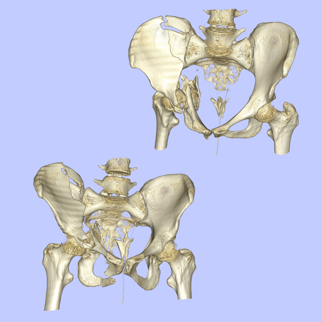

A case study: pre-operative and post-operative 3D models of a severe pelvic fracture.
I processed CT scan data through direct volume rendering to create 3D visualizations comparing the severe pelvic fracture before and after surgical intervention.


A case study: Human tooth
I created both direct volume rendering (left image) and indirect volume rendering (right image) of a human tooth to demonstrate different visualization approaches for dental anatomy.


A case study: Brain Tumour
This case involved working with both MITK and 3D slicer to generate brain tumour visualization from multi-modal imaging: MRI, CT scan, and PET scan data. I used direct volume rendering to create 3D visualization.
This approach demonstrates how multiple imaging modalities can be combined to provide tumour appearance for surgical planning and patient communication.


A case study: Lung Tumour
I used both direct and indirect volume rendering techniques to visualize a lung tumour case. These rendering techniques provide insights in density, location, shape, and volume of lung tumour.
The left image shows direct volume rendering, while the right image demonstrates indirect volume rendering of the same dataset.
All segmentations and visualizations were created in: