A Pilot’s Journey
Female reproductive systems are essentially understudied in marine mammals; however, understanding them is crucial for species conservation and reproductive health monitoring. Current documents, such as textbook photographs, histology, and ultrasound images, cannot entirely convey the complex 3D transformations that occur during reproductive cycles. Moreover, to imagine their anatomical changes through times.
For my master’s degree dissertation, I developed an interactive 3D web-based learning platform that allows learners to explore ovarian anatomical transformations in long-finned pilot whales through multiple reproductive stages, with integrated MRI comparisons, bilingual accessibility, and other features. This project is made possible by the support of my supervisors from the Glasgow School of Art & University of Glasgow, and a collaboration with Scottish Marine Animal Stranding Scheme (SMASS).
This tool serves as a prototype on how we can share the knowledge internationally and what we can contribute to marine biology and veterinary science education by combining conventional ways of learning with the use of technologies.
An Interactive 3D Web-Based Platform
The key point of this application is that users can interact with 3D anatomical models through a web platform. This tool transforms static scientific reading into visual, interactive education that can reach anyone and anywhere.
In the beginning of June 2025, I started exploring gaps in female reproductive biology study in marine mammals. While their reproductive system follow similar patterns to humans (yes! we are all mammals), a clear picture of the changes in female reproductive system remain limited.
The challenges I found can split into two areas:
Study limitations
Difficulty studying these animals in natural habitat and unpredictable wildlife encounters prevent long-term monitoring. Most knowledge comes from stranded individuals and necropsies.
Learning material gaps
Current materials rely heavily on 2D presentations (textbook photos, histology, ultrasound) that reduce 3D structures to 2D, losing spatial detail and changes over time. Not to mention new learners or someone who never experience this topic before, it is a hard subject.

These gaps and limitations highlighted the need for advanced imaging technique (And my supervisor, Mariel ten Doeschate, is currently doing research on this stuff!). Even though I couldn’t develop innovative medical imaging technology, I can develop a learning tool to help us understand this topic in greater depth.
That's when this prototype project was developed with collaborative supervision from the Glasgow School of Art, University of Glasgow and the Scottish Marine Animal Stranding Scheme (SMASS).
My sincere gratitude to my amazing supervisors:
Dr. Matthieu Poyade
Mariel ten Doeschate
Dr. Andrew Brownlow


Overview Session
“We all need a map before starting our great exploration. The overview is here at your service. ”
Design Behind Book
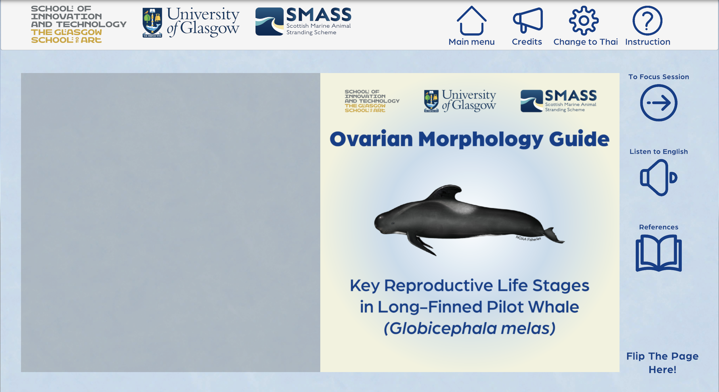

Traditional anatomy education in medical schools begins with illustrated textbooks. I transformed this familiar concept into an interactive design.
The overview session features a book-style interface that introduces basic concept of female reproductive biology in cetaceans, focusing on morphological changes in ovaries. Another features include voice-over narration and language switching, giving learners multiple ways to absorb information before entering the focus session.
Why long-finned pilot whales? The reason I chose this species because of a phenomenon we still don’t fully understand: post-reproductive life stage (or what we call “menopause” in female humans). One key question is that do long-finned pilot whales have a post-reproductive life stage (PRLS) like their relatives, short-finned pilot whales? The answer may lie within the ovaries. If we can read the story told by ovarian structures, we may unlock greater understanding of this phenomenon.
Fun fact! So far that we know, PRLS occurs in only a few species - humans, Asian elephants, and four cetaceans: beluga, narwhal, killer whale, and short-finned pilot whales (McCormack et al., 2023; Monaghan and Ivimey-Cook, 2023).
Focus Session
“To understand reproductive life, I think we need to see its changes not only from the outside, but also from within.”
Seeing the Internal


Histology image
showing cortex and medulla area of ovary

Illustration
showing cortex and medulla area of ovary
MRI image
showing cortex and medulla area of ovary

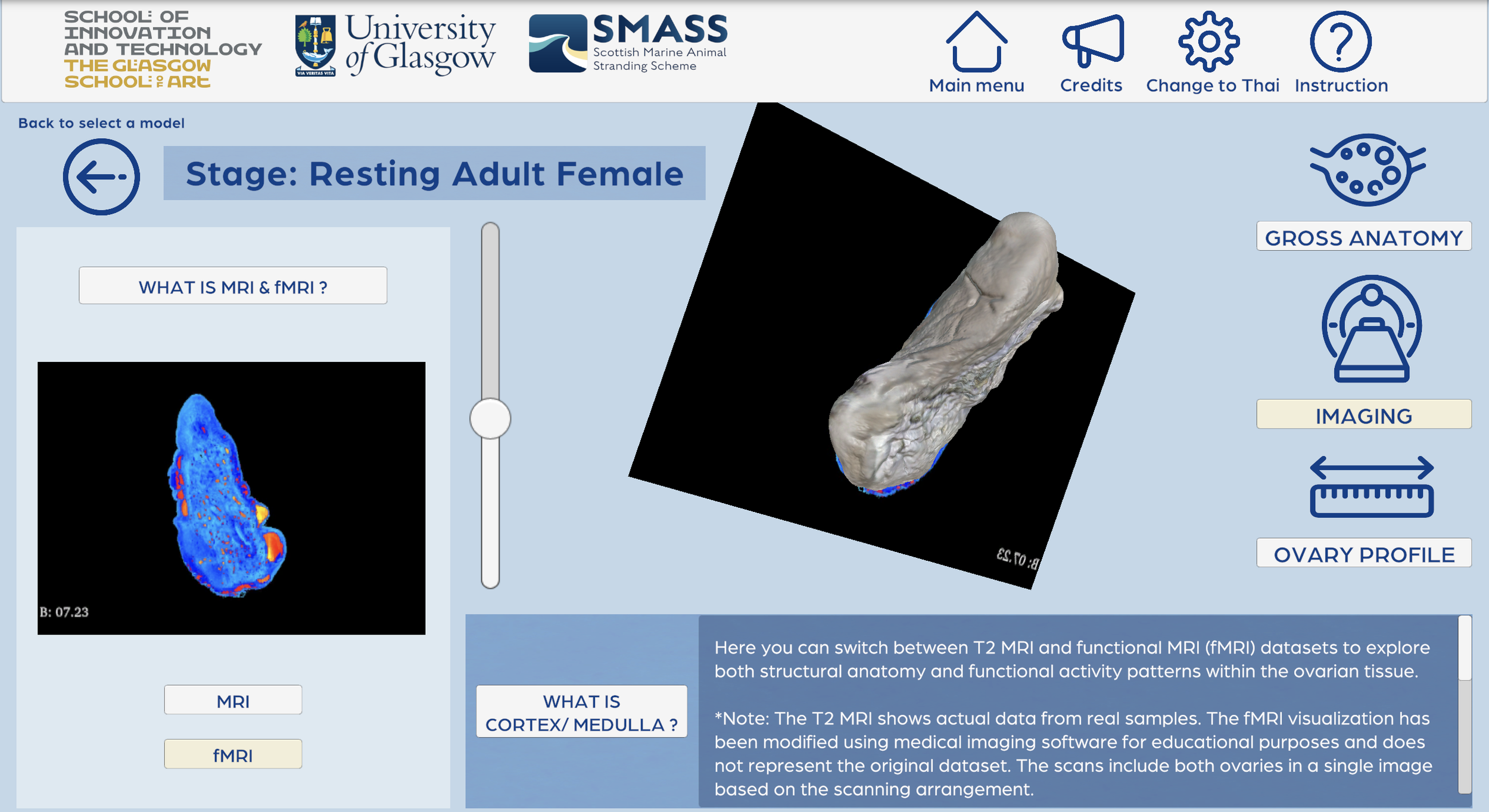
There are multiple ways to study internal structures of ovaries as you see the images above. Personally, the most challenging aspect was designing a way to present both internal and external ovarian structures simultaneously. With the help of my supervisors and MRI dataset of long-finned pilot whales’ ovaries, I developed an imaging panel feature that allows users to scroll through the 3D ovarian model while viewing corresponding MRI image stacks side-by-side.
Key innovation: Learners can switch between MRI and fMRI views. The original MRI dataset shows internal structures in grayscale. I adapted the fMRI color module in 3D Slicer to adjust the color palette of medical images. This visualisation highlights the differences between cortex and medulla areas of the ovaries, though further study is needed to fully validate this approach.
Other panels include Gross Anatomy and Ovary Profile, these serve learners to explore the 3D model in 360 degrees while comparing it corresponding with 2D photographs and also to access specimen specific details such as carcass conditions at time of death.
Note: The models do not depict natural colors due to the formalin fixation process used in specimen preservation.
Creating the 3D models


In part of ovarian models, I used 3D slicer, 3ds Max, and Zbrush.
My workflow include:
3D slicer: To segment MRI data as a 3D model file
3ds Max: To retopologize high-polygon models, making it clean and ready for developing forward in application
Zbrush: Final texture and color refinement for realistic visual appearance
This pipeline allowed me to create and capture every anatomical detail reliably from MRI data while creating models that work smoothly in the interactive platform.
Softwares used for creating 3D models:
Evaluation
“Simplicity and clarity”
“I like that you could see the 3D model and move through it to see the MRI at different stages, it was very visual. ”
“Easy to use”
For user testing and validation. 22 professionals participated in this study: 14 veterinary professionals, 4 marine mammal researchers, 1 marine biologist, and 3 from other related fields. All English and Thai-speaking professionals were included to test the application.
I used five metrics to evaluate the effectiveness of this application including knowledge assessment, cognitive load, face and content validity, usability, and users motivation.
Five Evaluation Metrics
Knowledge Acquisition
1. Knowledge Acquisition: Participants provided more correct answers after using the tool (pre-test mean score = 3.91, SD = 1.41 and post-test mean score = 4.59, SD = 0.66) and showed a statistically significant difference between pre- and post-test scores (Z = -2.652, p < 0.05).
Cognitive Loads
2. Cognitive Load: The application design properly managed mental effort required for learning complex anatomy. However, it is suggested that the design of application has meaningful impacts on their cognitive effort.
Face and Content Validity
3. Face and Content Validity: Participants showed moderate to positive perceptions of the 3D ovarian models’ realism and educational content quality.
Usability
4. Usability: The application achieved a SUS score of 75.57 (above the benchmark of 68). Users appreciated the ease of use and educational content quality.
Users Motivation
5. Users Motivation: Results indicated moderate to high motivation levels across all elements - attention, relevance, satisfaction, and confidence. Relevance scored lowest, showing variability in how participants perceived the tool’s applicability to their professional contexts.
Future Development: In brief, participants suggested valuable improvements including having juvenile and pregnant stage ovaries to complete the whole picture of female long-finned pilot whales reproductive cycle, histological/ultrasound images, inter-species comparison, dictionary pop-ups, and more gamification elements.
Conclusion: Overall this project presents positive results and reveals the need of further study and development of advanced learning materials for this field. Testing with broader and diverse audiences (such as veterinary students) is also needed to validate the effectiveness of this tool across experience levels.