


Creating 3D medical visualization by segmented and reconstructed Medical Imaging Data
Medical imaging captures incredibly detailed data about our bodies (both human and animal), but this information often remains in formats only medical professionals can interpret.
⎯ 𓁹 ⎯
ภาพถ่ายทางการแพทย์ทำให้เราเห็นภาพรายละเอียดของอวัยวะและชีวิตได้ชัดเจนขึ้น แต่บ่อยครั้งที่ข้อมูลเหล่านั้นถูกจำกัดอยู่ในรูปแบบที่ผู้เชี่ยวชาญเท่านั้นที่เข้าใจ การสร้างภาพ 3 มิติออกมาจะช่วยทำให้เราเห็นภาพชัดเจนยิ่งขึ้น
Seeing the inside
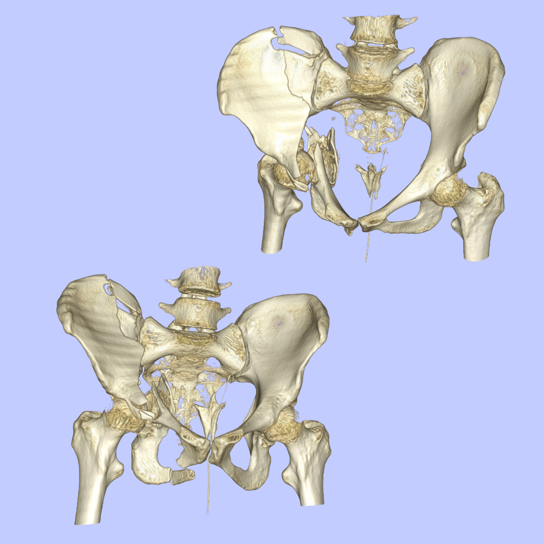

A case study: pre-operative and post-operative 3D models of a severe pelvic fracture.
I processed CT scan data through direct volume rendering to create 3D visualizations comparing the severe pelvic fracture before and after surgical intervention.
โมเดล 3 มิติ เพื่อเปรียบเทียบผลการผ่าตัดกระดูกเชิงกรานแตกหัก: ข้อมูลภาพถ่ายคอมพิวเตอร์ (CT Scan) ถูกนำมาผ่านกระบวนการ Direct Volume Rendering เพื่อสร้างภาพจำลอง 3 มิติ แสดงให้เห็นถึงความแตกต่างระหว่าง "ก่อน" และ "หลัง" การรักษา


A case study: Human tooth
I created both direct volume rendering (left image) and indirect volume rendering (right image) of a human tooth to demonstrate different visualization approaches for dental anatomy.
ภาพจำลองฟันมนุษย์แบบ 3 มิติ: สร้างขึ้นเพื่อเปรียบเทียบให้เห็นถึงความแตกต่างระหว่างสองเทคนิคหลักในการเรนเดอร์ข้อมูลทางการแพทย์ (direct volume rendering ภาพด้านซ้าย และ indirect volume rendering ภาพด้านขวา)


A case study: Brain Tumour
This case involved working with both MITK and 3D slicer to generate brain tumour visualization from multi-modal imaging: MRI, CT scan, and PET scan data. This approach demonstrates how multiple imaging modalities can be combined to provide tumour appearance for surgical planning and patient communication.
การรวมข้อมูลภาพถ่ายทางการแพทย์เพื่อจำลองเนื้องอกในสมอง 3 มิติ เคสนี้ผสานชุดข้อมูลจากหลากหลายแหล่ง (Multi-modal Imaging) ทั้ง MRI, CT และ PET Scan เพื่อสร้างภาพจำลองเนื้องอกขึ้นมา


A case study: Lung Tumour
I used both direct and indirect volume rendering techniques to visualize a lung tumour case. These rendering techniques provide insights into the density, location, shape, and volume of the lung tumour.
The left image shows direct volume rendering, while the right image demonstrates indirect volume rendering of the same dataset.
การจำลองภาพเนื้องอกในปอด งานนี้แสดงให้เห็นถึงความสำคัญของการเลือกใช้เทคนิคการเรนเดอร์ที่แตกต่างกัน เพื่อดึงข้อมูลสำคัญของเนื้องอกออกมาให้ได้มากที่สุด ทั้งในด้านความหนาแน่น ตำแหน่ง รูปร่าง และปริมาตร
All segmentations and visualizations were created in: