An Interactive 3D Web-Based Platform
แพลตฟอร์มการเรียนรู้ 3D แบบตอบโต้ได้บนเว็บไซต์
The key point of this application is that users can interact with 3D anatomical models through a web platform. This tool transforms static scientific reading into visual, interactive content that can reach anyone and anywhere.
หัวใจของการพัฒนาแอปพลิเคชันนี้คือการก้าวข้ามข้อจำกัดของการเรียนรู้แบบเดิม โดยการเปลี่ยนเนื้อหาวิชาการที่เคยอยู่แค่ในตำราให้กลายเป็นโมเดล 3 มิติที่โต้ตอบได้จริงบนเว็บไซต์ เครื่องมือนี้ช่วยสร้างประสบการณ์การเรียนรู้แบบใหม่ และสร้างพื้นที่เรียนรู้ออนไลน์ ที่ไม่ว่าใคร อยู่ที่ไหนในโลก ก็สามารถเข้าถึงความรู้ได้อย่างง่ายดาย !
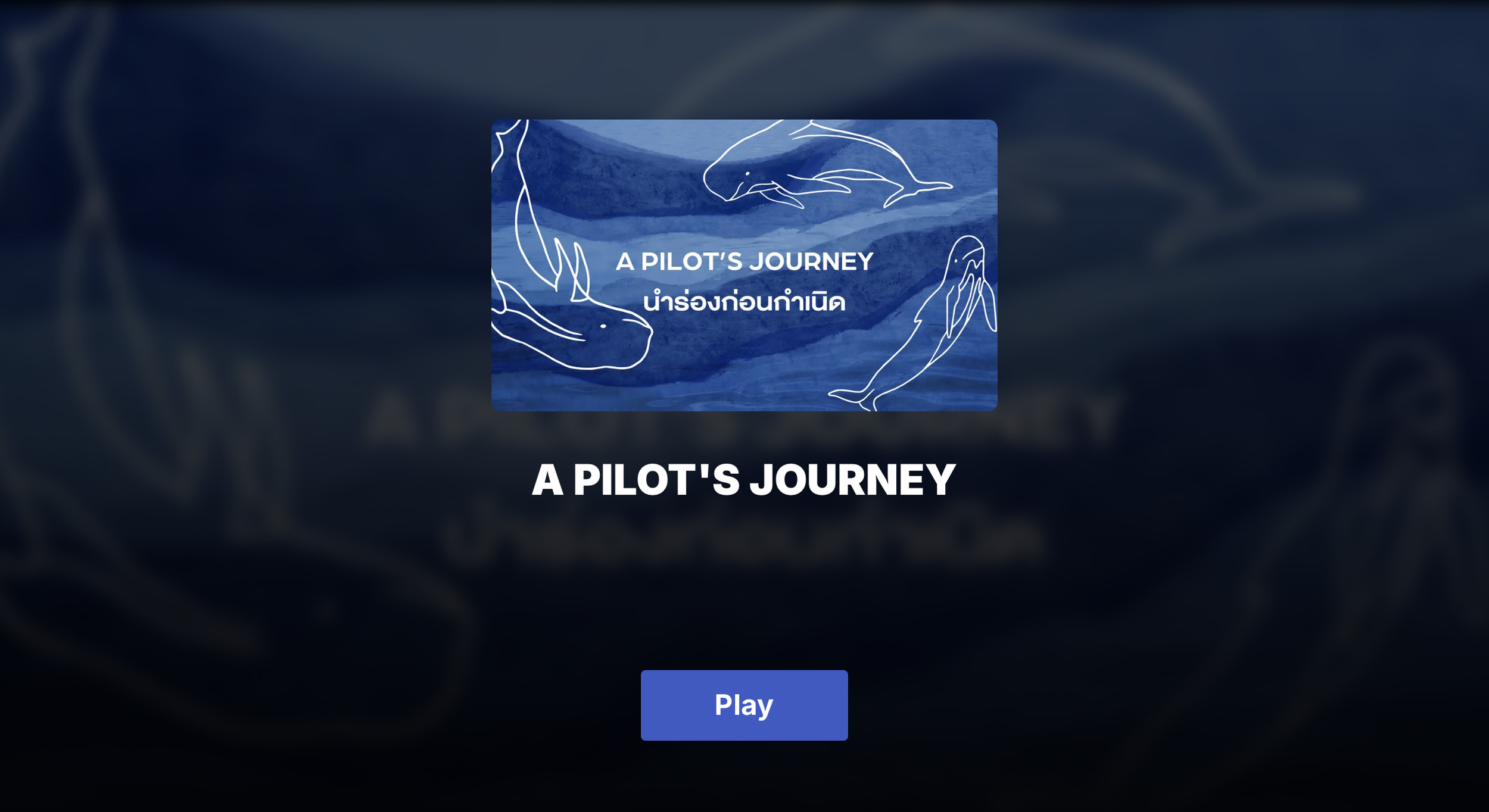
A Pilot’s Journey
นำร่องก่อนกำเนิด
⎯ 𓆝 𓆟 𓆞 𓆝 𓆟 ⎯
Female reproductive systems are essentially understudied in marine mammals; however, understanding them is crucial for species conservation and reproductive health monitoring. Current documents, such as textbook photographs, histology, and ultrasound images, cannot entirely convey the complex 3D transformations that occur during reproductive cycles. Moreover, to imagine their anatomical changes through time.
For my master’s degree dissertation, I developed an interactive 3D web-based learning platform that allows learners to explore ovarian anatomical transformations in long-finned pilot whales through multiple reproductive stages, with integrated MRI comparisons, bilingual accessibility, and other features. This project is made possible by the support of my supervisors from the Glasgow School of Art & University of Glasgow, and a collaboration with Scottish Marine Animal Stranding Scheme (SMASS).
This tool serves as a prototype for how we can share knowledge internationally and for what we can contribute to marine biology and veterinary science education by combining conventional learning methods with technology.
การศึกษาเกี่ยวกับระบบสืบพันธุ์เพศเมียในสัตว์เลี้ยงลูกด้วยนมทางทะเลยังไม่ค่อยมีรายงานออกมามากนักในทางวิชาการ อย่างไรก็ตามการศึกษาเพื่อให้เกิดความเข้าใจในระบบนี้มีความสำคัญมากในทางการอนุรักษ์และการเฝ้าติดตามสถานะสุขภาพการสืบพันธุ์ของสัตว์เลี้ยงลูกด้วยนมทางทะเล ปัจจุบันเรามีการบันทึกข้อมูลเกี่ยวกับระบบสืบพันธุ์ของสัตว์เพศเมียผ่านภาพในตำราเรียน ภาพทางจุลกายวิภาคศาสตร์ (histology) และภาพอัลตราซาวนด์ ซึ่งข้อมูลเหล่านี้ยังไม่สามารถที่จะนำเสนอความซับซ้อนของอวัยวะและการเปลี่ยนสภาพของเนื้อเยื่อในระหว่างวงจรการสืบพันธุ์ได้ครบทุกองค์ประกอบ
แพลตฟอร์มการเรียนรู้ 3D บนเว็บไซต์ (Interactive 3D Web-based Platform) จึงถูกพัฒนาขึ้นเพื่อให้ผู้เรียนได้สำรวจการเปลี่ยนแปลงของรังไข่วาฬนำร่องครีบยาว (Long-finned Pilot Whales) แบบรอบด้าน ผ่านโมเดล 3 มิติ ที่ถูกสร้างจากภาพ MRI จริงให้เห็นภาพชัดเจน พร้อมฟังก์ชันสลับได้ 2 ภาษา (ไทย-อังกฤษ) เพื่อการใช้งานในระดับสากล โดยโปรเจคดังกล่าวได้ถูกดำเนินการและให้คำปรึกษาจากทีมอาจารย์ที่ปรึกษาจาก the Glasgow School of Art & University of Glasgow และความร่วมมือจากทีม Scottish Marine Animal Stranding Scheme (SMASS)
เครื่องมือนี้เป็นตัวอย่างที่แสดงให้เห็นว่าเราสามารถนำเสนอข้อมูลทางวิชาการได้ชัดเจนยิ่งขึ้นผ่านการใช้สื่อภาพประกอบ รวมถึงสามารถใช้นวัตกรรมดิจิทัลมาช่วยในการสร้างเครื่องมือการเรียนรู้ทางสัตวแพทยศาสตร์หรือชีววิทยาทางทะเล เพื่อก้าวข้ามขีดจำกัดของการเรียนรู้แบบเดิม ที่ต้องผ่านตำราหรืออยู่แต่ในห้องเรียน ให้เข้าถึงง่ายขึ้นและมีความเป็นสากลมากยิ่งขึ้น
In early June 2025, I began exploring gaps in research on female reproductive biology in marine mammals. While their reproductive systems follow patterns similar to those of humans (yes! we are all mammals), a clear picture of the changes in the female reproductive system remains limited.
ต้นเดือนมิถุนายน 2025 อรเริ่มศึกษาค้นคว้าเพื่อหาช่องว่างทางการศึกษาด้านชีววิทยาการสืบพันธุ์เพศเมียในสัตว์เลี้ยงลูกด้วยนมทางทะเล แม้ว่าระบบสืบพันธุ์ของพวกเขาจะมีรูปแบบพื้นฐานคล้ายคลึงกับมนุษย์ แต่ภาพรวมของการเปลี่ยนแปลงในระบบนี้ยังมีข้อมูลรายงานอยู่น้อยมาก
⎯ ༄.°⎯
The challenges I found can be split into two areas:
Study limitations
Difficulty studying these animals in their natural habitat and unpredictable wildlife encounters prevent long-term monitoring. Most knowledge comes from stranded individuals and necropsies.
Learning material gaps
Current materials rely heavily on 2D presentations (textbook photos, histology, ultrasound) that reduce 3D structures to 2D, thereby losing spatial detail and temporal changes. Not to mention new learners or someone who has never experienced this topic before, it is a hard subject.
ความท้าทายในการศึกษาถูกแบ่งออกเป็น 2 ด้าน
ข้อจำกัดในการศึกษา: การศึกษาสัตว์ทะเลในถิ่นที่อยู่ตามธรรมชาติเป็นเรื่องยาก ความไม่แน่นอนของการพบเจอสัตว์ตามธรรมชาติทำให้การติดตามความเปลี่ยนแปลงในระยะยาวเป็นเรื่องท้าทาย ข้อมูลส่วนใหญ่จึงมักมาจากเคสสัตว์เกยตื้นหรือการชันสูตรเท่านั้น
สื่อการเรียนรู้: สื่อการสอนในปัจจุบันยังติดกับรูปแบบ 2 มิติ ซึ่งทำให้โครงสร้างที่มีความซับซ้อนแบบ 3 มิติถูกลดทอนรายละเอียดลงไป โดยเฉพาะเรื่องของมิติด้านพื้นที่ (Spatial detail) และการเปลี่ยนแปลงของโครงสร้างเนื้อเยื่อตามเวลา นักศึกษาสัตวแพทย์หรือผู้ที่ไม่เคยมีประสบการณ์ในด้านนี้มาก่อน อาจทำความเข้าใจได้ยาก


These gaps and limitations highlighted the need for an advanced imaging technique (and my supervisor, Mariel ten Doeschate, is currently researching this!). Even though I couldn’t develop innovative medical imaging technology, I can develop a learning tool to help us understand this topic in greater depth.
จากข้อจำกัดที่พบ การใช้เทคนิคการสร้างภาพวินิจฉัยขั้นสูง (Advanced Imaging) อย่างเช่น MRI เข้ามาช่วยอาจทำให้เห็นภาพการเปลี่ยนแปลงของโครงสร้างชัดเจนขึ้น แม้ว่าโปรเจคนี้จะไม่ได้มุ่งเน้นไปที่การพัฒนาระบบถ่ายภาพทางการแพทย์โดยตรง แต่เราสามารถสร้าง "เครื่องมือการเรียนรู้" ที่จะช่วยแปลงข้อมูลที่ซับซ้อนให้เข้าใจได้ง่ายและลึกซึ้งยิ่งขึ้นผ่านเทคโนโลยีในปัจจุบัน
That's when this prototype project was developed under collaborative supervision from the Glasgow School of Art, the University of Glasgow,and the Scottish Marine Animal Stranding Scheme (SMASS).
โปรเจคนี้ถูกสร้างขึ้นผ่านการดูแลจากทีมที่ปรึกษาจาก The Glasgow School of Art, University of Glasgow และ Scottish Marine Animal Stranding Scheme (SMASS) เพื่อผสานข้อมูลทางวิทยาศาสตร์และงานออกแบบดิจิทัลเข้าด้วยกัน
⎯ ༄.°⎯
My sincere gratitude to my amazing supervisors:
Dr. Matthieu Poyade
Mariel ten Doeschate
Dr. Andrew Brownlow


Overview Session
“We all need a map before starting our great exploration. The overview is here at your service.
เพราะทุกการเดินทางที่ยิ่งใหญ่ จำเป็นต้องมีแผนที่นำทางเสมอ!”
Behind the Design
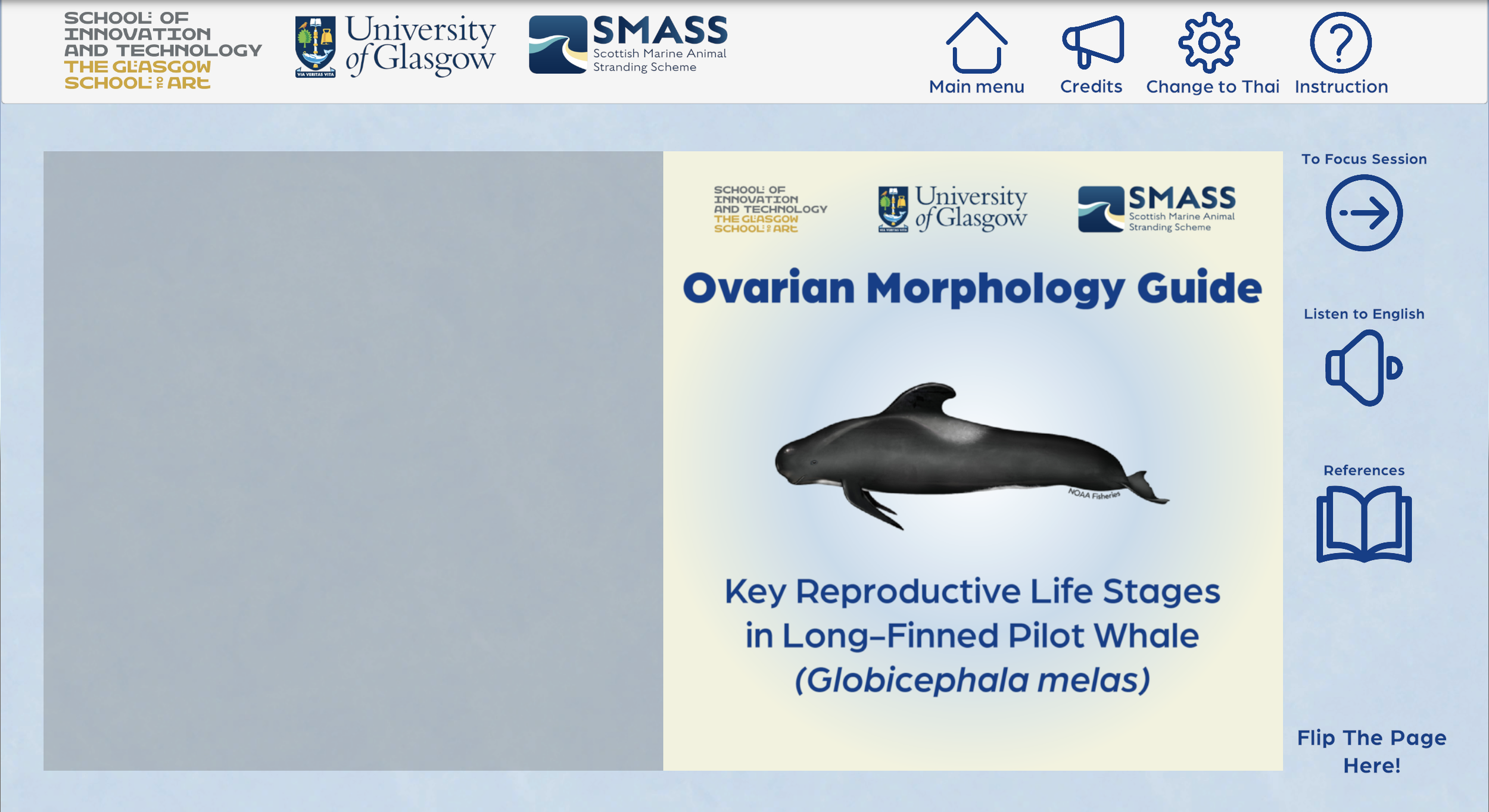

Traditional anatomy education in medical schools begins with illustrated textbooks. I transformed this familiar concept into an interactive design.
The overview session features a book-style interface that introduces basic concepts of female reproductive biology in cetaceans, focusing on ovarian morphological changes. Other features include voice-over narration and language switching, giving learners multiple ways to absorb information before entering the focus session.
มาเปลี่ยนตำราเรียนที่คุ้นเคย ให้กลายเป็นประสบการณ์ที่โต้ตอบได้ การเรียนกายวิภาคแบบดั้งเดิมมักเริ่มจากภาพวาดในตำรา อรจึงหยิบเอาคอนเซปต์ที่ทุกคนคุ้นเคยนี้มาแปลงโฉมใหม่ให้เป็นงานดีไซน์ที่โต้ตอบได้ โดยในส่วนของ Overview Session นี้ได้ถูกออกแบบในสไตล์ "สมุดภาพดิจิทัล" ที่เล่าเรื่องระบบสืบพันธุ์ของสัตว์เลี้ยงลูกด้วยนมทางทะเล พร้อมฟีเจอร์เสียงบรรยาย (Voice-over) และการสลับภาษา เพื่อให้ผู้เรียนได้ซึมซับข้อมูลพื้นฐานก่อนเข้าสู่การเรียนรู้เชิงลึก
⎯ ༄.°⎯
Why long-finned pilot whales? The reason I chose this species is a phenomenon we still don’t fully understand: the post-reproductive life stage (or what we call “menopause” in female humans). One key question is that do long-finned pilot whales have a post-reproductive life stage (PRLS) like their relatives, short-finned pilot whales? The answer may lie within the ovaries. If we can read the story told by ovarian structures, we may unlock a greater understanding of this phenomenon.
Fun fact! So far, we know that PRLS occurs in only a few species: humans, Asian elephants, and four cetaceans: beluga, narwhal, killer whale, and short-finned pilot whales (McCormack et al., 2023; Monaghan and Ivimey-Cook, 2023).
วาฬนำร่องครีบยาว มีเรื่องราวทางชีววิทยาที่นักวิทยาศาสตร์กำลังทำการศึกษา นั่นคือ “ภาวะหมดวัยเจริญพันธุ์ ” (หรือที่ในมนุษย์เรียกว่าวัยทอง) ซึ่งเป็นปรากฏการณ์ที่หาได้ยากในธรรมชาติ คำถามสำคัญคือ วาฬนำร่องครีบยาวมีช่วงชีวิตหลังหมดวัยเจริญพันธุ์เหมือนญาติสนิทอย่างวาฬนำร่องครีบสั้นหรือไม่? คำตอบอาจซ่อนอยู่ใน “รังไข่ ” หากเราสามารถ “ อ่าน ” เรื่องราวที่โครงสร้างรังไข่บอกเล่าได้ เราอาจจะไขความลับนี้ได้สำเร็จ
จนถึงปัจจุบัน เราพบภาวะหมดวัยเจริญพันธุ์ในสิ่งมีชีวิตเพียงไม่กี่ชนิดเท่านั้น ซึ่งได้แก่ มนุษย์, ช้างเอเชีย และวาฬอีก 5 สายพันธุ์ (วาฬเบลูก้า, วาฬนาร์วาล, วาฬเพชฌฆาต และวาฬนำร่องครีบสั้น) เท่านั้น!
“To understand reproductive life, I think we need to see its changes not only from the outside, but also from within.
เรื่องราวของชีวิต เพียงภายนอกอาจเล่าเรื่องราวไม่ครบทุกมุม...เพื่อให้เรื่องราวสมบูรณ์ อย่าลืมมองเข้าไปภายในด้วย”
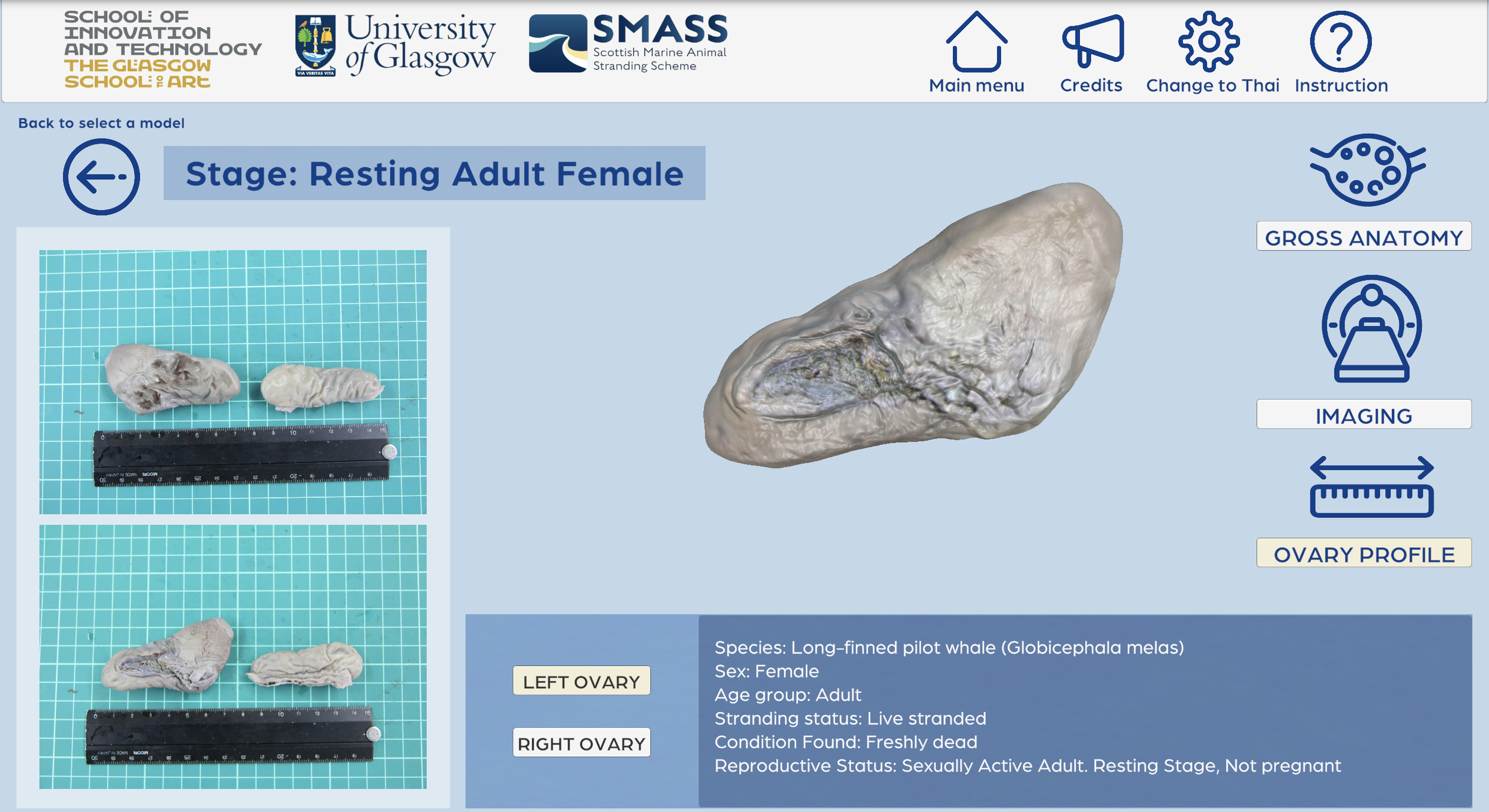
Focus Session
Seeing the Internal


Histology image
showing cortex and medulla area of ovary

Illustration
showing cortex and medulla area of ovary
MRI image
showing cortex and medulla area of ovary

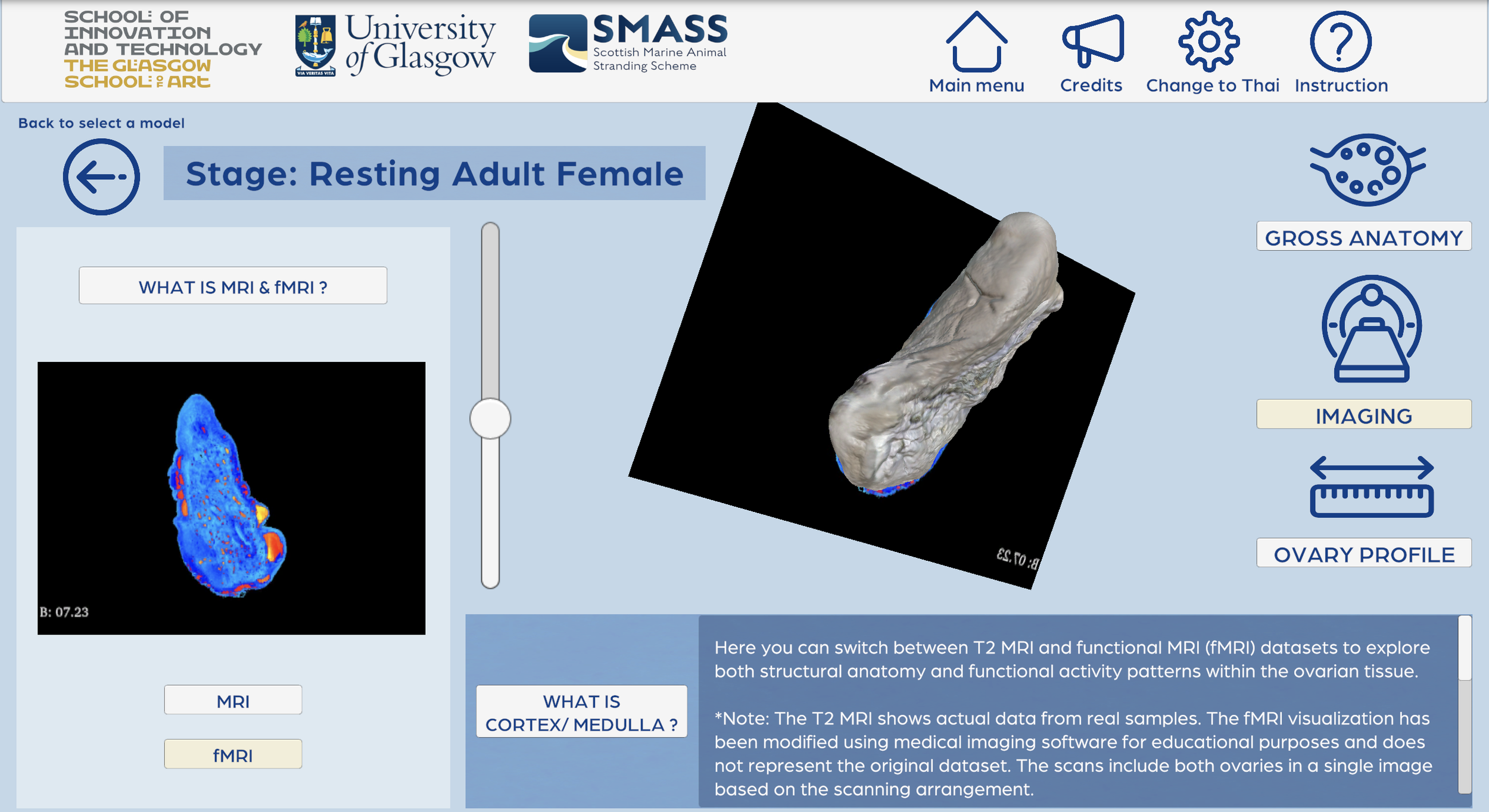
There are multiple ways to study internal structures of ovaries as you see the images above. Personally, the most challenging aspect was designing a way to present both internal and external ovarian structures simultaneously. With the help of my supervisors and MRI dataset of long-finned pilot whales’ ovaries, I developed an imaging panel feature that allows users to scroll through the 3D ovarian model while viewing corresponding MRI image stacks side-by-side.
Focus Session สร้างขึ้นมาเพื่อผสานมิติการมองเห็นให้ครบจบที่เดียว ความท้าทายที่สุดของโปรเจกต์นี้คือ จะทำอย่างไรให้ผู้เรียนเห็นทั้งโครงสร้างภายนอกและภายในได้พร้อมกัน "Imaging Panel" จึงถูกพัฒนาขึ้นมา เพื่อให้ผู้ใช้สามารถเลื่อนดูสแต็กภาพ MRI ควบคู่ไปกับการหมุนโมเดล 3 มิติแบบ Side-by-side ได้อย่างอิสระ
⎯ ༄.°⎯
Key innovation: To improve clarity, I introduced a toggle between standard MRI and an fMRI-inspired color module. While original MRI data appears in grayscale, I utilized 3D Slicer to adapt a color palette that highlights the contrast between the cortexand medulla areas. This visualization serves as a pioneering approach to identifying internal structures, providing a clearer roadmap for future validation.
Other panels include Gross Anatomy and Ovary Profile; these allow learners to explore the 3D model in 360 degrees, compare it with 2D photographs, and access specimen-specific details such as carcass condition at the time of death.
Note: The models do not depict natural colors due to the formalin fixation process used in specimen preservation.
เพื่อช่วยในการจำแนกเนื้อเยื่อ อรได้พัฒนาฟีเจอร์สลับโหมดระหว่าง MRI ปกติกับโหมดสีแบบ fMRI โดยนำชุดข้อมูลขาวดำมาปรับผ่านโมดูลสีในโปรแกรม 3D Slicer เพื่อเน้นให้เห็นความแตกต่างระหว่างพื้นที่ส่วน Cortex และ Medulla ของรังไข่ให้ชัดเจนยิ่งขึ้น ซึ่งนับเป็นการทดลองนำเสนอข้อมูลรูปแบบใหม่ที่ช่วยยกระดับการวิเคราะห์ให้เห็นภาพมากกว่าเดิม
นอกจากนี้ยังมีแผงควบคุมอื่นๆ เพื่อการเรียนรู้ เช่น Gross Anatomy: สำรวจโมเดล 3 มิติได้ 360 องศา พร้อมเปรียบเทียบกับภาพถ่ายจริง และ Ovary Profile: เข้าถึงข้อมูลเฉพาะของแต่ละเคส เช่น สภาพของซากสัตว์ ณ เวลาที่พบ เพื่อประกอบการศึกษาเชิงลึก
หมายเหตุ: สีของโมเดล 3 มิติ เป็นสีที่เกิดจากกระบวนการรักษาสภาพตัวอย่างด้วยฟอร์มาลิน (Formalin Fixation) ไม่ใช่สีธรรมชาติของอวัยวะในขณะที่มีชีวิต
Creating the 3D models


In part of the ovarian models, I used 3D Slicer, 3ds Max, and ZBrush.
My workflow includes:
3D slicer: To segment MRI data as a 3D model file
3ds Max: To retopologize high-polygon models, making them clean and ready for developing in application
ZBrush: Final texture and color refinement for a realistic visual appearance
This pipeline allowed me to reliably capture every anatomical detail from MRI data while creating models that run smoothly on the interactive platform.
ในการสร้างโมเดลรังไข่จากตัวอย่าง MRI ให้ถูกตามหลักกายวิภาค กระบวนการทำงาน (Workflow) จึงถูกออกแบบขึ้นโดยผสานจุดเด่นของ 3 โปรแกรมหลัก (3D Slicer, 3ds Max, and ZBrush) เพื่อเปลี่ยนข้อมูล MRI ที่ซับซ้อนให้กลายเป็นโมเดลที่ใช้งานได้จริงบนเว็บไซต์
Softwares used for creating 3D models:
Results & Evaluation
“Simplicity and clarity
เรียบง่ายและชัดเจน”
“I like that you could see the 3D model and move through it to see the MRI at different stages, it was very visual.
งานดูสมจริง ชอบที่สามารถมองเห็นภาพโมเดล 3 มิติ และเลื่อนผ่านโมเดลเพื่อมองภาพ MRI ในแต่ละระยะได้”
“Easy to use
ใช้งานง่าย”
For user testing and validation. 22 professionals participated in this study: 14 veterinary professionals, 4 marine mammal researchers, 1 marine biologist, and 3 from other related fields. All English and Thai-speaking professionals were included to test the application.
เพื่อให้มั่นใจว่าแพลตฟอร์มนี้สามารถใช้งานได้จริงในเชิงวิชาการ เราได้เชิญผู้เชี่ยวชาญรวม 22 ท่าน มาร่วมทดสอบและประเมินผล ซึ่งประกอบด้วย: สัตวแพทย์ 14ท่าน นักวิจัยสัตว์เลี้ยงลูกด้วยนมทางทะเล 4 ท่าน นักชีววิทยาทางทะเล 1 ท่าน และผู้เชี่ยวชาญในสาขาที่เกี่ยวข้องอีก 3 ท่าน ทั้งกลุ่มผู้เชี่ยวชาญที่ใช้ภาษาไทยและภาษาอังกฤษ
⎯ ༄.°⎯
I used five metrics to evaluate the effectiveness of this application, including knowledge assessment, cognitive load, face and content validity, usability, and user motivation.
สำหรับโปรเจคนี้ ใช้เกณฑ์การประเมิน 5 ด้านหลัก เพื่อวัดผลสัมฤทธิ์ของแอปพลิเคชัน ประกอบด้วย 1)การประเมินความรู้ (Knowledge Assessment) เพื่อวัดความเข้าใจหลังการใช้งาน 2)ภาระทางปัญญา (Cognitive Load) เพื่อตรวจสอบว่าเนื้อหาไม่อัดแน่นจนเข้าใจยากเกินไป 3)ความตรงตามเนื้อหาและลักษณะภายนอก (Face & Content Validity) เพื่อการันตีความถูกต้องแม่นยำทางวิชาการของโมเดล 4)ความยากง่ายในการใช้งาน (Usability): เพื่อประเมินความลื่นไหลของระบบ Web Interface และ 5)แรงจูงใจของผู้ใช้ (User Motivation) เพื่อวัดว่าเครื่องมือนี้ช่วยกระตุ้นความยากเรียนรู้ได้มากน้อยเพียงใด
Five Evaluation Metrics
Knowledge Acquisition
1. Knowledge Acquisition: Participants provided more correct answers after using the tool (pre-test mean score = 3.91, SD = 1.41 and post-test mean score = 4.59, SD = 0.66) and showed a statistically significant difference between pre- and post-test scores (Z = -2.652, p < 0.05).
ผลการทดสอบแสดงให้เห็นว่า แพลตฟอร์มนี้ช่วยให้ผู้เข้าร่วมทดสอบมีความรู้ความเข้าใจเพิ่มขึ้นอย่างมีนัยสำคัญ โดยคะแนนแบบทดสอบหลังการใช้งานเพิ่มสูงขึ้นกว่าก่อนใช้งานอย่างเห็นได้ชัด
คะแนนเฉลี่ยก่อนทดสอบ (Pre-test): 3.91 (SD=1.41)
คะแนนเฉลี่ยหลังทดสอบ (Post-test): 4.59 (SD=0.66)
นัยสำคัญทางสถิติ: Z=−2.652, p<0.05
ตัวเลขเหล่านี้พิสูจน์ว่า เครื่องมือนี้มีประสิทธิภาพสูงในการถ่ายทอดความรู้ทางวิชาการที่ซับซ้อน
⎯ ༄.°⎯
Cognitive Loads
2. Cognitive Load: The application design properly managed the mental effort required for learning complex anatomy. However, it is suggested that the design of application has meaningful impacts on their cognitive effort.
ผลการประเมินพบว่า แอปพลิเคชันนี้สามารถจัดการ Cognitive Load ได้อย่างเหมาะสม แม้ว่าเนื้อหาทางกายวิภาคจะมีความซับซ้อนสูง แต่เครื่องมือเรียนรู้ของโปรเจคนี้ถูกออกแบบมาเพื่อทำให้ผู้เรียนไม่รู้สึกว่าต้องใช้ความพยายามมากเกินไปในการทำความเข้าใจ
วิเคราะห์ได้ว่า งานดีไซน์มีผลกระทบโดยตรงต่อประสิทธิภาพการเรียนรู้ การจัดระเบียบข้อมูลที่ดีช่วยให้ผู้เรียนสามารถจดจ่อกับการทำความเข้าใจเนื้อหาได้อย่างเต็มที่ โดยไม่ถูกขัดจังหวะด้วยความซับซ้อนของตัวแอปพลิเคชัน
⎯ ༄.°⎯
Face and Content Validity
3. Face and Content Validity: Participants showed moderate to positive perceptions of the 3D ovarian models’ realism and educational content quality.
ผู้เข้าร่วมทดสอบมีความคิดเห็นในเชิงบวกอย่างมากต่อ ความสมจริง (Realism) ของโมเดลรังไข่ 3 มิติ และคุณภาพของเนื้อหาที่นำเสนอ โดยผลประเมินอยู่ในเกณฑ์ ดีมาก ผลการประเมินนี้ยืนยันว่า งานดีไซน์ของโมเดลไม่ได้มีดีแค่ความสวยงาม แต่ยังมีความถูกต้องแม่นยำตามหลักกายวิภาคศาสตร์ ซึ่งเป็นหัวใจสำคัญในการนำไปใช้เป็นสื่อการเรียนรู้ที่เป็นมาตรฐานสากล
⎯ ༄.°⎯
Usability
4. Usability: The application achieved a SUS score of 75.57 (above the benchmark of 68). Users appreciated the ease of use and educational content quality.
จากการประเมินด้วยระบบ SUS (System Usability Scale) แพลตฟอร์มนี้ได้รับคะแนนสูงถึง 75.57 ซึ่งสูงกว่าค่าเฉลี่ยมาตรฐานสากลที่ 68 ผู้ทดสอบต่างให้การยอมรับว่าตัวแอปพลิเคชันใช้งานง่าย (Ease of use) และมีการจัดวางเนื้อหาทางวิชาการที่มีคุณภาพได้อย่างลงตัว
⎯ ༄.°⎯
Users Motivation
5. User Motivation: Results indicated moderate to high motivation levels across all elements - attention, relevance, satisfaction, and confidence. Relevance scored lowest, indicating variability in participants' perceptions of the tool’s applicability to their professional contexts.
ผลการประเมินในด้านแรงจูงใจผ่านเกณฑ์มาตรฐานในระดับ “กลาง-สูง" ในทุกองค์ประกอบ (ARCS Model) ซึ่งสะท้อนถึงการสร้างประสบการณ์การเรียนรู้ที่น่าสนใจ; Attention & Satisfaction: ผู้ใช้ให้ความสนใจและมีความพึงพอใจต่อรูปแบบการนำเสนอ; Confidence: ระบบ Interactive ช่วยสร้างความมั่นใจให้ผู้เรียน ว่าสามารถทำความเข้าใจเนื้อหาที่ยากได้จริง; Relevance หรือการประยุกต์ใช้: ในส่วนนี้มีความคิดเห็นที่หลากหลาย ซึ่งถือเป็นข้อมูลที่มีค่ามาก เนื่องจากช่วยชี้ให้เห็นว่า การนำเครื่องมือนี้ไปใช้ควรปรับให้เหมาะกับบริบทของแต่ละวิชาชีพ (เช่น งานสัตวแพทย์ภาคสนาม กับงานวิจัยในห้องแล็บ) ที่มีความต้องการแตกต่างกันออกไป
⎯ ༄.°⎯
Future Development: In brief, participants suggested valuable improvements, including having juvenile and pregnant stage ovaries to complete the whole picture of female long-finned pilot whales' reproductive cycle, histological/ultrasound images, inter-species comparison, dictionary pop-ups, and more gamification elements.
เพื่อให้เครื่องมือนี้เป็นสื่อการสอนที่สมบูรณ์ยิ่งขึ้น ควรเพิ่มเติมในเรื่องของโมเดลรังไข่ในระยะวัยเด็ก (Juvenile) และระยะตั้งท้อง (Pregnant) เพื่อให้เห็นภาพรวมของวงจรการสืบพันธุ์ที่ครบถ้วน /เพิ่มการเปรียบเทียบกับภาพทางจุลกายวิภาค (Histology) และอัลตราซาวนด์ เพื่อการวิเคราะห์ที่แม่นยำขึ้น/ เพิ่มฟีเจอร์เปรียบเทียบระหว่างสายพันธุ์ เพื่อเพิ่มความเข้าใจในเชิงวิวัฒนาการ และสุดท้ายเพิ่มพจนานุกรมคำศัพท์เฉพาะทางแบบ Pop-up รวมถึงองค์ประกอบแบบเกม (Gamification) เพื่อให้การเรียนรู้สนุกและสามารถจดจำได้แม่นยำยิ่งขึ้น!
⎯
Conclusion: Overall, this project presents positive results and reveals the need for further study and development of advanced learning materials for this field. Testing with broader, diverse audiences (such as veterinary students) is also needed to validate the tool's effectiveness across experience levels.
สรุป โปรเจกต์นี้เป็นต้นแบบที่พิสูจน์ให้เห็นถึงพลังของเทคโนโลยีดิจิทัลในการยกระดับการเรียนรู้เกี่ยวกับวิทยาศาสตร์ทางทะเล แม้ผลตอบรับจะเป็นไปในทิศทางที่ดี แต่ก็ควรทำการทดสอบกับกลุ่มผู้ใช้งานที่หลากหลายขึ้น เช่น นักศึกษาสัตวแพทย์ เพื่อปรับปรุงให้เครื่องมือนี้มีประสิทธิภาพ เหมาะสำหรับผู้เรียนในทุกระดับ และสามารถใช้เครื่องมือนี้เป็นส่วนหนึ่งในการขับเคลื่อนการศึกษาและเรียนรู้ด้านการอนุรักษ์สัตว์น้ำในระดับสากลต่อไป